Inspire Roadshow brings Life and Health Sciences alive for local primary students

24 students from a local South Bristol primary school were captivated by a Roadshow visit of four staff and postgraduate students in November 2018.

The Year 6 students had the opportunity to do a carousel of 4 different interactive activities designed to complement their recent science lessons about healthy bodies and lifestyle. Staff from the Dental Hospital ran an interactive station about the formation of plaque and the importance of oral hygiene. The students loved getting hands on cleaning bacteria from the giant teeth with a giant toothbrush!

A PhD student from the Clinical Nutrition research group, led a healthy eating station where the student enjoyed learning the basis of good nutrition from the healthy eating mat.

A PhD student from the Bristol Platelet Group, built on the student’s knowledge of heart rates and human anatomy. They enthusiastically worked through the body parts apron, learning about each organ in turn. They also had the chance to use pulse oximeters to measure their own heart rate and oxygen levels. The students enjoyed testing the effect of exercise on their heart rate.

On the final station, the students were taught the basics of CPR using the Annie resus models and were able to have a go themselves. They remarked how hard CPR is and how tired they became from just a short time but how they would be able to have a go if needed or help someone else understand what to do.

Zebrafish: The new scientific superhero

The use of zebrafish in research by MSc student Alistair Vorster

The zebrafish (Danio rerio) is becoming an increasingly valuable organism for scientists studying the human genome and various disorders. This little freshwater fish may represent one of the most important tools for future research into developmental biology, and has already helped reveal a number of key genes associated with various human disorders, including those affecting blood cells, the heart and kidneys (e.g. bradycardia and congenital anaemia) (1).

Zebrafish are a tropical fish found in South East Asia, and their use in research began in the 1960’s (2). They have several characteristics which make them ideal for scientific research, including rapid reproduction and the ability to produce large numbers of transparent larvae which develop in eggs fertilised outside the womb, allowing any changes to be observed easily during early stages of development. They are also easy to look after, requiring less space than rodents and exhibiting quicker development than their mammalian counterparts. Their entire genome has been sequenced, with the latest update released in 2017 (3), which is of great use for genetic research through comparison with the human genome. 70% of human genes have a zebrafish counterpart, with 84% of human disease associated genes displaying a zebrafish ‘sibling’ (4). Genetic pathways and genes for development are often very similar between humans and zebrafish, such as for the heart, therefore they are an excellent model for vertebrate genetics and development.

Superhero powers?

One characteristic of zebrafish which makes them particularly interesting is their ability to regrow damaged tissue, including heart muscle, fin tissue and even neurons (5). This phenomenon, reminiscent of lizards that escape predators by losing their tail which then miraculously regrows, is leading scientists to investigate the methods of regeneration used by the zebrafish. It is hoped that by revealing the biological secrets behind this process we may be able to manipulate human cellular mechanisms to repair damaged tissue in the future, e.g. for sufferers of heart disease.

Research has shown that a special group of cells, called the blastema, is important for this regeneration. These cells form a bundle at the point of injury and show characteristics similar to stem cells, meaning that they can create the different types of tissue needed for regrowth (6). Interestingly, mature bone cells have also been found to revert back into more immature ‘stem’ like cells after injury and during regrowth, meaning that they can help to make new bone e.g. during fin regrowth (7).

Research at Bristol

Research groups at the University of Bristol have been carrying out studies using zebrafish to help better understand their regenerative abilities. Dr Chrissy Hammond leads a group focusing on research examining the regulation of cartilage and bone homeostasis in development and in disease, whilst Dr Beck Richardson’s work focuses on inflammatory signalling pathways and how this affects skin and cardiac repair. Together these groups have helped publish new work into the regenerative mechanisms of zebrafish. During the last few months at the University of Bristol, I have been involved in research for my masters degree working with both Dr Hammond’s and Dr Richardson’s groups, using zebrafish to examine regeneration of the tail fin and the factors involved.

Previous studies have suggested that the immune system may play an important role in regeneration, therefore a major focus of the project was observing immune cell behaviour. This was done using fluorescent fish lines (called transgenics) and immunostaining (which uses antibodies to label specific cell types) to visualise individual immune cells after injury to the tail fin. Genetic manipulation means that transgenic fish can produce immune cells of certain fluorescent colours, which can be examined by microscope to track their behaviour during regeneration.

An exciting result from the research found a mutant fish line may have a less mature immune system, which may in turn make that fish less effective at regenerating tissue. Fish were subjected to gene ‘knock out’, whereby particular genes are lost and therefore these fish lack certain proteins. Immunostaining showed that mutants without the gene in question have smaller and fewer immune cells compared to normal zebrafish. This represents a good example of how zebrafish can help to identify key factors involved in regeneration and disease. Previous research has also identified mutants such as sau, which affects a gene responsible for blood cell development. Humans lacking this gene exhibit a type of hereditary anaemia, therefore this zebrafish mutant can be used as a model for this disease (1), and highlights the capacity for zebrafish to replace other lab animals, e.g. rodents, and take a more prominent role in future medical research.

The future of medical research?

Just how important could zebrafish be for future medical science? Given their suitability as a vertebrate research model, I believe the zebrafish will be an increasingly used tool in future medical studies. Zebrafish can benefit various disease models including cancer studies, as shown by researchers in Lisbon who have been using zebrafish to track tumour growth and examine treatment with different experimental drugs (8). Tumours marked in fluorescence can be tracked in these fish at a quicker and cheaper rate than with rodents, and cancers have been shown to respond and behave in the same way in the fish as in humans.

And it is not only in academia where zebrafish are popular. Pharmaceutical companies such as Roche consider them a valuable research tool that can increasingly replace rodents in their studies, made easier due to recent advances in genetic manipulation (9). Researchers are also increasingly aware of and striving towards the 3 R’s, refinement, reduction and replacement of animals. The 3 R’s aim to find ways of using alternatives to animals in research, reduce the numbers of animals used in each study and refine methods to reduce pain and suffering experienced by research animals (more details can be found on the NC3R website https://www.nc3rs.org.uk/the-3rs). Increasing use of zebrafish over ‘higher’ vertebrate species such as rodents would represent a step in the right direction, with potentially less opposition to their use in scientific research.

I must admit that when I began my project with these creatures I was not aware of their abilities and potential to contribute to scientific research. However, having seen their regenerative capacities in real time and the crucial work that the research groups at the university are carrying out, I believe that they can be a great tool for clinical research that will be increasingly used going forward. I hope that new studies, including a recently funded programme which will see zebrafish sent into space to examine physiological changes at zero gravity, will shed new light on vertebrate biology and add to the growing importance of these scientific superheroes.

References
1. Dooley K, Zon LI. Zebrafish: a model system for the study of human disease. Curr Opin Genet Dev. 2000;10(3):252-6.
2. Unknown. Why use the zebrafish in research 2014 [Available from: https://www.yourgenome.org/facts/why-use-the-zebrafish-in-research.
3. Consortium GR. Zebrafish Genome 2017 [Available from: https://www.ncbi.nlm.nih.gov/grc/zebrafish.
4. McKie R. How the diminutive zebrafish is having a big impact on medical research The Guardian2013 [Available from: https://www.theguardian.com/science/2013/sep/15/zebrafish-human-genes-project.
5. Poss KD, Wilson LG, Keating MT. Heart Regeneration in Zebrafish. Science. 2002;298(5601):2188.
6. Poss KD, Keating MT, Nechiporuk A. Tales of regeneration in zebrafish. Developmental Dynamics. 2003;226(2):202-10.
7. Knopf F, Hammond C, Chekuru A, Kurth T, Hans S, Weber Christopher W, et al. Bone Regenerates via Dedifferentiation of Osteoblasts in the Zebrafish Fin. Developmental Cell. 2011;20(5):713-24.
8. Leslie M. Zebrafish implanted with a cancer patient’s tumor could guide cancer treatment: Science; 2017 [Available from: http://www.sciencemag.org/news/2017/08/zebrafish-implanted-cancer-patient-s-tumor-could-guide-cancer-treatment.
9. Roche. Zebrafish Roche; 2018 [Available from: https://www.roche.com/research_and_development/drawn_to_science/zebrafish.htm.

Antimicrobial resistance in Thailand: a holistic approach

Antimicrobial resistance (AMR) is one of the most serious global threats to human health in the 21st century. One of the Bristol researchers taking on this challenge is Professor Matthew Avison who is leading the ‘One Health Drivers of Antibacterial Resistance in Thailand’ consortium project, supported by the Medical Research Council. Here, he tells us about the benefits of working together across borders and disciplines, and how the consortium’s approach can help inform AMR research worldwide.

In Thailand, AMR is estimated to have led to 38,000 deaths in 2010 and cost the economy $1.2 billion. Since then, the problem has continued to grow.

The Thai authorities are monitoring the situation closely and the World Health Organisation recognises their surveillance as an exemplary model for other low and middle income countries (LMICs). But the research to date has been in discrete areas.

This is making the key biological, social, cultural and economic drivers behind AMR (and how they all interact) hard to pinpoint. The long-term impacts of Thailand’s educational campaigns and regulations aimed at curbing AMR are also largely unknown, making effective evidence-based preventative measures difficult to design.

A gut problem

Two of the deadliest bacterial species that have acquired antibiotic resistance are Klebsiella pneumoniae (K. pneumoniae) and Escherichia coli (E. coli), and they’re the ones our ‘One Health’ project focuses on.

They live in the guts of all mammals and we’re interested in how and why they might spread between animals and humans, through the food chain and the environment. Their prevalence in human infections in Thailand has been rapidly increasing. And, worryingly, infections caused by antibiotic-resistant K. pneumoniae have risen from none before 2010, to 10-20% in 2016.

Covering all AMR bases

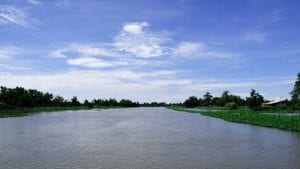
Our project takes a more holistic view of this complex problem. We’ve chosen an 80km square region in the central and western area of Thailand for our study: the Mae Klong-Ta Chin Basin. We picked it due to its incredible diversity of landscapes, populations and land use.

There are small villages, large towns and industrial zones, with communities of varying socio-economic and education levels. It includes fish farms, fruit orchards and rice paddies. Such variation is crucial because it gives us the opportunity to investigate and compare potential drivers of AMR across different cultural and environmental settings.

And our team is just as varied. We’ve brought together a consortium of UK and Thai researchers covering a wide spectrum of specialities, including veterinarians, clinicians, ecologists, chemists, microbiologists, mathematicians, statisticians, social scientists and project managers. Each researcher benefits from having a mirror version of themselves based in the other country, to collaborate with and learn from.

Operation data collection

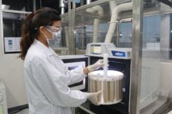
Our data will cross disciplines, from chemical analysis of environmental and food samples, to findings of anthropological studies into how and why people use antibiotics. Over three years, we’ll repeat our collection cycles at specific sites and in different weather conditions and seasons.

Then our mathematicians will help us understand the patterns and relationships within this data. And our timing coincides with a Thai review into their National Strategic Plan on AMR, so we hope our findings will help influence future policy that will benefit people in Thailand.

Local and global connections

We’ve found that collaborating with on-the-ground Thai researchers who are connected to the local healthcare and farming systems is invaluable. They help engage with people in communities with varied levels of poverty and literacy and negotiate the sensitive ethical and practical considerations of data collection.

Involving early-career researchers from both countries has also been a huge benefit. They’ve brought fresh ideas, new ways of working and an energy that’s revitalised our approach. I hope this early experience of international collaboration will encourage and support them to continue in academic research.

We live in a small, connected world and the devastating impact of AMR stretches all around it. Our hope is that our project can complement work being undertaken in other communities and countries. AMR is such a multi-layered issue with huge ramifications across all aspects of human society. Our findings and those of our peers can help meet this global challenge and contribute to maintaining food security, combating poverty, reducing pollution and protecting human health.


 ‘One Health Drivers of Antibacterial Resistance in Thailand’ It is a consortia project between the Chulabhorn Research Institute, Mahidol University, University of Bristol, University of Bath, University of Exeter and the NERC Centre for Ecology and Hydrology.

 This project is one of four ‘AMR in a Global Context’ Consortia awards, totalling £12 million, which have been jointly funded by the cross-research council AMR initiative and the National Institute for Health Research (NIHR).

 All four projects will contribute to the UK’s commitment to Official Development Assistance (ODA).  The Research Councils’ contribution will be made through the Global Challenges Research Fund (GCRF) which supports cutting edge research addressing the problems faced by developing countries. The NIHR contribution supports human health research and will be made through its Global Health Research ODA allocation. NIHR’s ODA funding is aimed at supporting internationally-outstanding applied research for the direct and primary benefit of patients and the public in low and middle-income countries (LMICs).


Engineering Red Blood Cells

PhD student Joseph Hawksworth describes his work developing tools to genetically engineer red blood cells

Genetically edited BEL-A cells under the microscope

My last three years have been spent engineering red blood cells, working towards the ultimate goal of production of customised red blood cells for therapeutic uses. I’m a final year PhD student studying Synthetic Biology at the University of Bristol and I work in Dr Ashley Toye’s group which specialises in cultured blood in the laboratory. There are many definitions of Synthetic biology but common to them all is an emphasis on the design and engineering of biological systems for useful purposes. In the spirit of this, we aim to produce ‘designer’ red blood cells (RBCs) to help overcome numerous challenges, for example replicating disease states for study, improving properties of lab grown blood, or even utilising RBCs as drug delivery vehicles. This month my work was published in EMBO Molecular Medicine (DOI: 10.15252/emmm.201708454). Here I’ll give some background to the project and describe the challenges, failures, and successes along the way!

In order to produce designer RBCs, we needed to develop a reliable way of editing the genome. Fortunately, I joined the lab at an opportune moment as two exciting technologies were emerging: CRISPR-Cas9 gene editing and a new RBC precursor cell line called BEL-A (Bristol Erythroid Line – Adult). CRISPR-Cas9 technology was being hailed as a revolutionary new approach to gene editing, with many laboratories reporting successful edits in a variety of cell types. CRISPR-Cas9 consists of two components: Cas9, a protein which cuts DNA, and a guide RNA which binds to Cas9 and directs it to the target DNA site. Once DNA has been cut it can be repaired, either through faulty DNA repair, preventing gene function (knockout), or through repair using a third component, template DNA, which enables very precise edits of the DNA sequence. Whilst CRISPR gene editing was gaining in popularity, my co-supervisor, Dr Jan Frayne, was developing the BEL-A cell line which offered an endless supply of RBC precursor cells. The BEL-A cells offered an excellent testbed for CRISPR based editingand the goal of my PhD was to bring together these two nascent technologies.

Multiple blood group proteins were targeted for removal

Encouraged by CRISPR success stories from laboratories worldwide, we started with an ambitious attempt to insert a large piece of DNA into the genome. Unfortunately this was unsuccessful, so we opted for a simpler approach, creating gene knockouts. We tested a variety of ways for getting the CRISPR components into the BEL-A cells and found a lentiviral approach to be most efficient. Initially, genes associated with membrane proteins were targeted, such as glycophorin A, as these were easiest to detect presence/absence from the cell surface. This time the editing worked! We were excited at the implications; most blood groups are determined by membrane proteins (or sugars attached to these proteins), therefore the removal of membrane proteins potentially enables us to make RBCs with increased transfusion compatibility.

It can be extremely difficult to source blood for transfusions of people with very rare blood types. Likewise, people with blood diseases, such as sickle cell anemia and thalassemia, often require repeated blood transfusions with difficult-to-source blood which has been carefully matched to minor blood group antigens in order to avoid adverse transfusion reactions. We wanted to target proteins which, if removed, would theoretically serve the broadest possible transfusion requirement. To identify a ‘hit list’ of proteins for removal, we collaborated with Dr Fiona Regan (NHSBT Hammersmith) who conducted a 15-month NHSBT survey to identify which antigens most commonly caused difficulty in matching blood for transfusions.The blood group proteinsRh, Kell, Duffy and GPB, as well as the ABO group (a carbohydrate), were identified as key targets. Using CRISPR-Cas9 we were able to generate BEL-A lines deficient for each of these targets and confirmed that the cells were still capable of developing into reticulocytes (young RBCs).

A dish of ‘Multi-Compatible BEL-A’ cells

The next stage was to combine all of the above blood group knockouts in a single cell to produce a cell with a theoretically enhanced transfusion compatibility. We completed several rounds of gene editing until we had knocked out all targets. One multiple knockout cell line was selected and we showed that it was possible to make reticulocytes which looked identical and had very similar properties to the original cells. The whole genome was sequenced and we found no extra mutations caused by the CRISPR-Cas9 edits, other than the ones we expected to observe. This is the first RBC precursor line developed which is absent for all of these blood group proteins and it was intriguing to see that these absences could be tolerated in reticulocytes derived from this cell line.These cells could theoretically serve 48 out of the 56 cases identified in the initial survey,representing a significant step towards the ability to tailor blood groups to meet the needs of patients!

Despite the significant progress we’ve made in developing engineered RBCs, there remain a number of challenges before translating our work to a clinical scenario. The work has been completed on a laboratory scale, and the numbers of cells we have been working with would require significant scale up. Similarly, the current costs of cell culturing are prohibitively expensive for therapeutic use. Finally, the cell line is currently not approved for use in humans, and overcoming this would require the production of clinical-grade cells produced according to ‘good manufacturing practice’ requirements. However, this is important proof-of-concept work which demonstrates the potential for designer RBCs developed for therapeutic use in humans. The first recipients of such cells would likely be people who are difficult, if not impossible, to match blood for transfusion requirements at present. The next stage of this work for the Toye lab is developing more advanced CRISPR methods allowing greater control over the genome and this is what I’m currently working on.

On a personal note, I feel extremely lucky to publish the first paper of my PhD on such an exciting and fast-moving project. I am grateful to the publication’s co-authors, in particular Tim Satchwell who I worked closely with, and all members of the Toye laboratory, for their insight, support and collaboration. Now to write my thesis!

55 Yr 6 and Yr 9 students have fun at Southmead

I have just returned from 2 amazing days of Primary and Secondary taster sessions being held at Southmead Hospital on the 4th and 5th of July.  The event put on by the NHS Clinical Teaching Fellows and the University of Bristol with 55 students from 6 different local WP schools attending the event.

“hits home as to why I got involved with outreach as a career”

The day was designed to inspire, inform and motivate students and help them understand more about the different careers related to the healthcare and health science professions.  The interactive session really helped open the students eyes to the range of roles within this healthcare sector as they met and spoke with doctors, HCA’s and patients.

The students clearly gained a lot from the day as beatifically summed up by a letter Mohammed wrote after the event.


I think this event has really helped Mohammed and reading his letter really hits home as to why I got involved with outreach as a career.


The students loved getting hands on experience of Basic Life Support as they practised resuscitation and the Recovery Position.  They learnt about the lungs by measuring their peak flow and listening to them through stethoscopes.  They discovered all about the heart by measuring their own electrical activity on the ECG machine,  handled a plasticised ox heart, measured their pulse and oxygen saturation levels and saw how they changed with exercise.  They decided that it was handy to know all this before their Sports Day the following day!

They also had a guided tour of the hospital and met the wounded lion sculpture outside the main Brunel Building.  They had a talk from some doctors, HCA’s and patients about their experiences working and using the NHS.

The event coincided with the 70th anniversary of the NHS and the students were able to witness lots a celebratory activity going on in the main atrium from cake sales to piano playing and raffles.

The whole day was nicely captured by this pupil’s drawing of the healthcare professionals she has heard about during the day that she presented to the Clinical Fellows to put up on their coffee room wall.

LGBT STEM day

On Thursday 5th July we celebrated LGBTSTEM day, the first International Day of LGBTQ+ People in Science, Technology, Engineering and Maths.

The campaign is organised by four groups: Pride in STEM, House of STEM, InterEngineering, and Out in STEM; and is supported by over 40 organisations including CERN, the European Space Agency, and Wellcome. A day of recognition honours the memory of LGBTQ+ scientists who came before us and supports those who are currently in STEM. We believe it should be possible to follow your passion whilst being yourself.

We celebrated LGBTSTEM Day in Biochemistry with a rainbow themed coffee and cake morning, and also collected donations for a local charity which works with LGBTQ+ young people called Off The Record. Off The Record is a mental health charity working across Bristol and South Gloucestershire. Off The Record run a number of LGBTQ+ youth groups and offer one-to-one counselling with LGBTQ+ young people – all of their services are free of charge.

The official hashtag for LGBTSTEM day is #LGBTSTEMDay.

Bristol University cleaner Herman Gordon cries tears of joy after students raise £1,500 for him to go on holiday

Herman Gordon, one of the most positive and friendly characters within the Biomedical Sciences Building was presented with the money and a note that said:

“On behalf of the students at Bristol, we would like to thank you for all the positive energy you have given to us throughout the years. You have brightened many of our days and we want you to know that we love and appreciate you. We have come together to give you a special gift as our way of saying thank you. Have a lovely summer!”

Wiping away his tears with his cleaning cloth Herman said:

“God bless you man, God bless, I want to say thank you for everything you have done and God bless every one of you.”

The students who helped raise the money chose to remain anonymous but issued a public statement:

“We would like to thank everyone who contributed to this incredible gift. It is truly amazing what we can achieve when we come together. It is thanks to your generosity that we’ve been able to raise about £1,500 – meaning both Herman and his wife will be able to visit Jamaica for the first time in 4 years!”

Creative reactions 2018 – Merging research and art

‘Creative Reactions’ brings together scientific researchers and artists to bring to life the amazing diversity of University research through the medium of art.

  • 'Hepatic' by Emily Krainc, a piece focused on research of Professor John Iredale based on fibrosis, the process of scarring.

Over 30 University of Bristol researchers from many different Schools got involved in Creative Reactions, including those from BiochemistryBiological SciencesCellular and Molecular Medicine and Experimental Psychology which from August this year will be part of the new Faculty of Life Sciences.

The event was a great success, with art being displayed in Hamilton House for members of the public view and talk to the researchers and artists behind each piece.

One of the event organisers Matthew Lee said:

‘Creative Reactions allows for engagement between researchers, artists and the public to allow researchers to communicate what their research is all about.

This is a great way to encourage the public to get involved with science and also encourages the academics and artists involved to think about their work from different perspectives.’

Event Organisers from left to right: Luke McGuinness, Charlotte Mugliston, Andrea Diaz-Gaxiola, Catherine Gilmore, Anouk Spelt and Matthew Lee.

Success at the Southwest Big Bang Fair 2018

The Southwest Big Bang Fair which ran on the 27 June is one of the biggest outreach events in the Southwest region, giving children the opportunity to engage with a range of different science exhibitors.

This year outreach volunteers from the upcoming Faculty of Life Sciences ran stands to highlight and enthuse thousands of school children, inspiring the next generation of scientists, who will be needed to help tackle global issues.


Dr Gail Born, Life Sciences Faculty Engagement Officer said:

“The Big Bang events are a wonderful opportunity to engage with and inspire thousands of primary and secondary students from around the southwest.

They are fully hands on and the students love being able see and use some amazing bits of equipment and talk to real scientists.”

The event is part of a programme of events taking place across the South West region, click here for more information on events near you.

Any staff or students interested in particpiating in future outreach events please contact Dr Gail Born

outreach-biomedsci@bristol.ac.uk