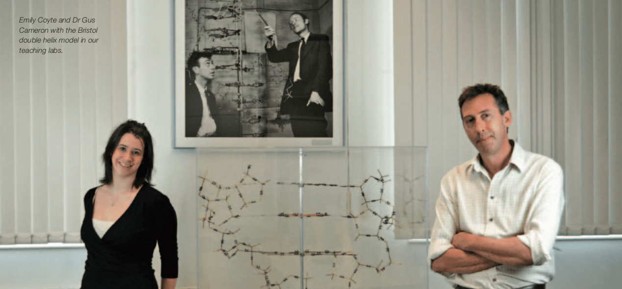
On this day in 1875 [9 June], Physiologist & Pharmacologist and Nobel Prize winner Henry Dale, was born. From failed pregnancies to wound repair, we spoke to two Sir Henry Dale Fellows in the School of Biochemistry about their research and how the fellowship for outstanding scientists is enabling them to answer some of life’s biggest questions.
Life’s great mysteries: Dr Binyam Mogessie
All human life starts with the fertilisation of an egg by a sperm. Numerous life processes that are invisible to the naked eye need to happen accurately, but unfortunately these tend to fail very often for reasons we are still trying to understand.
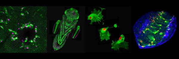
In my lab, we use very advanced microscopes to help us look inside mammalian eggs so that we can learn all the secrets behind how these giant cells eventually become a human being. To observe eggs in action, we use fluorescent molecular colours to label their DNA. We also label the tiny molecular machines that carry this DNA and the tracks they follow to move it from one location to another inside these large cells. We combine all of these to shoot spectacular and colourful movies of the life of an egg as it prepares itself to be fertilised by a sperm. We recently discovered that a protein called ‘Actin’ protects eggs from conditions that cause pregnancy failures and Down’s syndrome.
‘We recently discovered that a protein called ‘Actin’ protects eggs from conditions that cause pregnancy failures and Down’s syndrome.’
Since joining Bristol Biochemistry in 2018, I’m using powerful microscopes and even newer methods of studying proteins inside mammalian eggs. These advanced systems and many other research technologies in Bristol will accelerate discoveries that will help us in the future to protect human eggs from mistakes that prevent the birth of healthy babies. Being part of the vibrant School of Biochemistry means I get to work with some of the most talented young minds in the country. My long-term goal is to translate my research discoveries into treatments of human infertility and prevention of embryo deaths. This will ensure that the research we do in the lab today will ultimately have meaningful socio-economic impact that will improve human lives worldwide.
Research in my lab is supported by funding from the Wellcome Trust and the Royal Society through a Sir Henry Dale Fellowship. This generous funding has allowed us to acquire world-class imaging technologies and other tools needed to study mammalian egg development at unprecedented details. My Sir Henry Dale Fellowship has also helped me immensely in forming strong national and international scientific collaborations that continue to increase the impact of our research.
Why time doesn’t heal all wounds: Dr Helen Weavers
Our ability to regenerate, repair or ‘heal’ ourselves after injury is crucial as it seals the broken barrier and stops infection. However, unlike superheroes like Deadpool or Spider-Man, us humans (in most cases) can’t heal ourselves perfectly – we suffer problems, such as scar tissue, and for many people (including the elderly or diabetics) some wounds never heal.
‘Unlike superheroes like Deadpool or Spider-Man, us humans (in most cases) can’t heal ourselves perfectly – we suffer problems, such as scar tissue, and for many people (including the elderly or diabetics) some wounds never heal.’
In my research, I want to understand more about how our wounds heal – what do our individual cells do and which genes are involved – to help find ways we can improve the healing process. And for this, I use the fruit-fly! This might seem a surprising choice – why use an insect found in fruit bowls at home, to understand how human bodies work? But in fact, the not-so-humble fruit-fly has underpinned ground-breaking biomedical research for over a century and contributed to 5 Nobel Prizes! Fruit-flies and humans share many common organs (including a functional immune system) and 75% of human disease-causing genes. Fruit-flies are also optically translucent at many life-stages, which means we can image these fascinating processes – including the repair of wounds and recruitment of immune cells (‘inflammation’) – in real-time as they are happening live inside the fly, using fluorescently-tagged proteins to label different cell types. Fruit-flies are also ‘genetically tractable’ – meaning we can easily manipulate or ‘mutate’ individual genes to find out the gene’s function.
Bristol has been an ideal place for beginning my independent research career. It has state-of-the-art imaging equipment and brand-new facilities for breeding the flies! I’m also collaborating with researchers in a different department (‘Population Health Science’) to use real data from the human genome to help find genes important for wound repair.

Sir Henry Dale Fellowships, a partnership between the Royal Society and the Wellcome Trust, are for outstanding post-doctoral scientists wishing to build their own UK-based, independent research career addressing an important biomedical question.
It provides support for postdoctoral researchers who aim to become independent scientists leading their own groups.